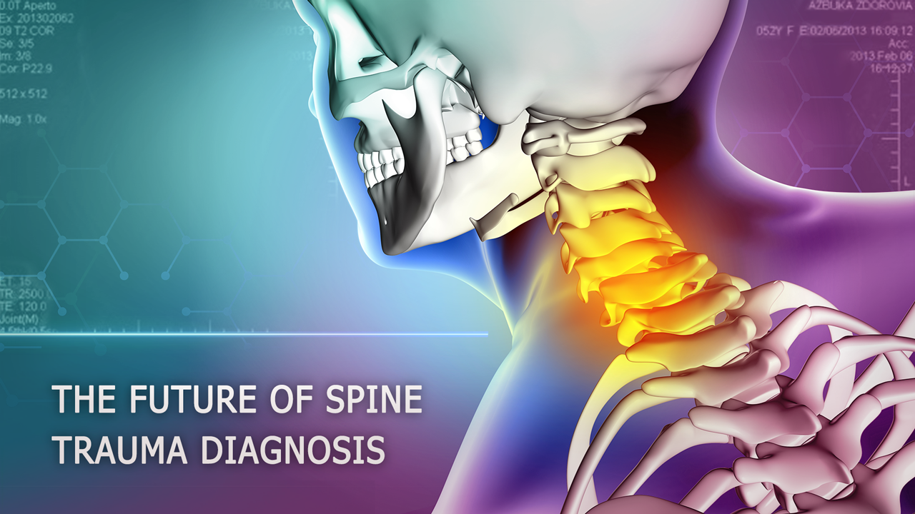
The Future of Spine Trauma Diagnosis: Advanced AI Techniques for Fracture Detection
Spinal injuries are some of the most devastating injuries a person can suffer. Among the most common spinal fractures is the cervical spine fracture, which can lead to paralysis and other serious complications if not identified and treated promptly. Unfortunately, detecting this type of fracture is often difficult, especially in elderly patients who may have degenerative diseases or osteoporosis that can mask the injury.
Fortunately, advances in machine learning technology are making it easier to identify spine fractures with greater accuracy and efficiency than ever before. By using a machine learning model, it is possible to train the system to recognize patterns and characteristics that may not be easily visible to the naked eye. This can lead to better accuracy in identifying the presence and location of cervical spine fractures, even in cases where it may be challenging due to age-related degenerative changes.
Machine learning is a form of artificial intelligence (AI) that allows systems to learn and improve from experience without being explicitly programmed. By analyzing large amounts of data, a machine learning model can learn to recognize patterns and make predictions based on that information.
In this article, we will explore how the Qudata team used machine learning to develop a highly accurate model for detecting cervical spine fractures from computed tomography (CT) scans.
The challenge of cervical spine fracture detection was presented in an open competition on Kaggle. The competition provided a platform for researchers and healthcare professionals to come together and create innovative solutions to the challenge of detecting spinal fractures. The competition also helped to highlight the importance of early detection and treatment of spinal fractures, as well as the potential of AI and machine learning in the field of medical imaging.
To prevent neurologic deterioration and paralysis after trauma, quickly detecting and determining the location of any vertebral fractures is essential. While previously techniques using X-ray radiation (X-rays) were used, imaging diagnosis of adult spine fractures is now almost exclusively performed with CT. One of the significant reasons for using CT instead of X-rays in spinal trauma is the speed with which the fracture site can be identified, minimizing or even preventing post-traumatic neurological complications or paralysis.
To solve the problem of cervical spine fracture detection, a ground truth dataset was created by a challenge planning task force. The dataset included imaging data sourced from twelve sites on six continents, including approximately 3,000 CT studies. Radiology specialists from the American Society of Neuroradiology (ASNR) and the American Society of Spine Radiology (ASSR) provided expert image level annotations for these studies to indicate the presence, vertebral level, and location of any cervical spine fractures.
As a starting point, a database of 2,000 patients with approximately 200 CT scans per person was processed. Due to the initial expert support provided by spine radiology specialists, the QuData team identified 1,000 people with fractures. Despite the large number of available CT scans for every patient with fractures, only a few of the images showed any fractures, and even those tended to be barely visible.

To avoid overfitting, the cervical spine vertebrae set was split into separate ones and worked individually, as well as with the full set. To achieve this, our team applied 2D semantic segmentation using vertebra masks provided only for 87 patients. Despite this limitation, the QuData team achieved an IoU metric* of approximately 0.98 after a few days of training on the Tesla T4**, indicating a high accuracy of the developed model, matching the ground truth dataset.

Overall, the detection of spinal fractures remains a significant challenge for healthcare providers. However, with the creation of ground truth datasets and the development of AI and machine learning models, there is hope for improving the accuracy and efficiency of spinal fracture detection. As the field of medical imaging continues to evolve, it is exciting to see how these technologies will shape the future of healthcare and contribute to better patient outcomes.
Read our case study Cervical spine fracture detection to learn more about the pipeline and technological details of the model provided by QuData.
*IoU metric – Intersection over Union metric is used in the image segmentation task and evaluates the segmentation quality. IoU shows how well the described object boundary matches the ground truth data.
Calculation of the IoU metric is performed by calculating the ratio of the area of intersection between the ground truth and the boundary obtained by the segmentation algorithm to the area of their union.
IoU = (Area of Overlap) / (Area of Union - Area of Overlap)
The IoU metric takes values from 0 to 1, where 1 means that the ground truth coincides with the boundary of the object selected by the segmentation algorithm. The closer the IoU value is to 1, the better the quality of the segmentation task performed.
** Tesla T4 is a graphics accelerator developed by Nvidia for data processing, machine learning and artificial intelligence. It is used to solve large computational problems.
Tesla T4 is one of the fastest and most efficient graphics cards for big data and machine learning.
Iryna Tkachenko, marketing manager
